
Thursday, November 29, 2012
Thursday, November 15, 2012
Final Observation
November 15, 2012
Last week a food pellet was inserted into my microaquarium, and this week I got to see it's effect.
Amount of Life vs. Diversity of Life : Results of the food pellet and of the microaquarium over time.
From last week to this week the amount of life increased greatly, but not nessecarly the diversity in life. But not that the diversity did not increase at all, I did find a few more additions that I had not seen before.
 (Forest 1954)
Another type of Green Algea I found hanging around. He moved kind of slow and squiggly like. He is multicellular. He is a Chlorophyll green, and I only saw a couple like him. They were spread throughout the glass.
(Forest 1954)
Another type of Green Algea I found hanging around. He moved kind of slow and squiggly like. He is multicellular. He is a Chlorophyll green, and I only saw a couple like him. They were spread throughout the glass.
 (Patterson 2003)
I found multiple little swarms of these Paramecium throughout the glass. This particular swarm was near the top and horizontally in the middle. They swim and run around in every which direction.They are single celled Protista and there were planty of them throughout the glass.
(Patterson 2003)
I found multiple little swarms of these Paramecium throughout the glass. This particular swarm was near the top and horizontally in the middle. They swim and run around in every which direction.They are single celled Protista and there were planty of them throughout the glass.
 (Patterson 2003)
This is the top veiw of a Stentor. I found two of these in my glass. It is funnel shaped and has cilia that causes the water and particles around it to swirl.He is unicellular.
(Patterson 2003)
This is the top veiw of a Stentor. I found two of these in my glass. It is funnel shaped and has cilia that causes the water and particles around it to swirl.He is unicellular.
 (Smith 2001)
I only found one of him. He was kicking around in some sediment on the right bottom of the glass. He is a good size and he is multicellular.
(Smith 2001)
I only found one of him. He was kicking around in some sediment on the right bottom of the glass. He is a good size and he is multicellular.
Along with these new additions, I observed some organisms already pictured and identitfied before, but they were in much greater quantity and, in the case of the cyclops, they were bigger in size.
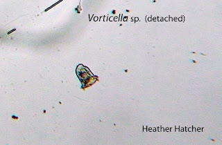
There were lots of tiny, unidentifed organisms swimming about also. There were about a dozen fairly large sized cyclops, three immature cyclops, and three dead ones.Oddly enough, most of them were in the left side of the glass. There were plent of rotifers throughout, I counted about twenty five before I gave up. I counted about twenty vorticellas throughout, but they were always in small groups. For the first time, I saw about half a dozen of them swimming around, not anchored to anything(pictured below).
 There was planty of green algea out and about, including the big blob mentioned last week. It has become a busy neighborhood. There was also more of it throughout the glass.

 Along with these lovely critters, there were about a half dozen seed shrimp (copadods) running around.
 Well my microaquarium has certainly been an adventure! Isn't it pretty? I am going to miss it.
I will post the final organisms totals later in a different post.
Last week a food pellet was inserted into my microaquarium, and this week I got to see it's effect.
Amount of Life vs. Diversity of Life : Results of the food pellet and of the microaquarium over time.
From last week to this week the amount of life increased greatly, but not nessecarly the diversity in life. But not that the diversity did not increase at all, I did find a few more additions that I had not seen before.
 (Forest 1954)
 (Patterson 2003)
 (Patterson 2003)
 (Smith 2001)
Along with these new additions, I observed some organisms already pictured and identitfied before, but they were in much greater quantity and, in the case of the cyclops, they were bigger in size.
There were lots of tiny, unidentifed organisms swimming about also. There were about a dozen fairly large sized cyclops, three immature cyclops, and three dead ones.Oddly enough, most of them were in the left side of the glass. There were plent of rotifers throughout, I counted about twenty five before I gave up. I counted about twenty vorticellas throughout, but they were always in small groups. For the first time, I saw about half a dozen of them swimming around, not anchored to anything(pictured below).


I will post the final organisms totals later in a different post.
Monday, November 12, 2012
Week 3 Observations
November 8, 2012 I went and observed my Microaquarium. A food pellet was added that morning before my observations.
FOOD PELLET INFORMATION:"Atison's Betta Food" made by Ocean Nutrition, Aqua Pet Americas, 3528 West 500 South, Salt Lake City, UT 84104. Ingredients: Fish meal, wheat flower, soy meal, krill meal, minerals, vitamins and preservatives. Analysis: Crude Protein 36%; Crude fat 4.5%; Crude Fiber 3.5%; Moisture 8% and Ash 15%.(McFarland)
So there were alot more organisms this week than last, in variety and in number. I also began to observe different algea. I found some new organisms this week too, and here is their photographs.
 (Pennak 1989)
 (Forest 1954)
One of the algea found in the glass. He and his algea friends seemed to be plentyful, which is odd because I didnt see a single on last week. They move around, but this particular one wasnt moving very fast.
(Forest 1954)
One of the algea found in the glass. He and his algea friends seemed to be plentyful, which is odd because I didnt see a single on last week. They move around, but this particular one wasnt moving very fast.
 (Pennak 1989)
He is kind of cute. He moves like a little tiny spider, and I saw a handful of others like him in the glass.
(Pennak 1989)
He is kind of cute. He moves like a little tiny spider, and I saw a handful of others like him in the glass.
 (Forest 1954)
These particular euglena were in one big blob together (last photograph) along with other organisms. They are some of my new found algea friends, and there seems to be lots of them. This particular blob of the was found in the bottom left of the tank, but I found some scattered throughout.
(Forest 1954)
These particular euglena were in one big blob together (last photograph) along with other organisms. They are some of my new found algea friends, and there seems to be lots of them. This particular blob of the was found in the bottom left of the tank, but I found some scattered throughout.
 (Forest 1954)
 (Patterson 2003)
Lots and lots of these swimming everywhere, and kind of in little groups. They seem unicellular and there is plenty of them.
(Patterson 2003)
Lots and lots of these swimming everywhere, and kind of in little groups. They seem unicellular and there is plenty of them.

I also observed alot of other organisms that images' have been previously posted. I saw about four copapods, an epistylelis, a cyclops, about thirty rotifers, several pinnularia near the left bottom of glass, several tiny organisms about, One big group of euglena as well as some scattered about, several trachelomas in one area close to the bottom left of the glass, a nematode, a meridion, five vorticellas, and a healthy amount of colpidium and other small organisms.Most of the life was in the bottom left of the glass, but there was still organisms spread about. Alot of the smaller ones grouped together.
FOOD PELLET INFORMATION:"Atison's Betta Food" made by Ocean Nutrition, Aqua Pet Americas, 3528 West 500 South, Salt Lake City, UT 84104. Ingredients: Fish meal, wheat flower, soy meal, krill meal, minerals, vitamins and preservatives. Analysis: Crude Protein 36%; Crude fat 4.5%; Crude Fiber 3.5%; Moisture 8% and Ash 15%.(McFarland)
So there were alot more organisms this week than last, in variety and in number. I also began to observe different algea. I found some new organisms this week too, and here is their photographs.
 (Pennak 1989)
 (Forest 1954)
 (Pennak 1989)
 (Forest 1954)
 (Forest 1954)
 (Patterson 2003)
I also observed alot of other organisms that images' have been previously posted. I saw about four copapods, an epistylelis, a cyclops, about thirty rotifers, several pinnularia near the left bottom of glass, several tiny organisms about, One big group of euglena as well as some scattered about, several trachelomas in one area close to the bottom left of the glass, a nematode, a meridion, five vorticellas, and a healthy amount of colpidium and other small organisms.Most of the life was in the bottom left of the glass, but there was still organisms spread about. Alot of the smaller ones grouped together.
Monday, November 5, 2012
Week 2 Observations
My microaquarium was observed on November 1, 2012
On Friday October 26, 2012 ONE Beta Food Pellet was inserted into the microaquarium.
FOOD PELLET INFORMATION:"Atison's Betta Food" made by Ocean Nutrition, Aqua Pet Americas, 3528 West 500 South, Salt Lake City, UT 84104. Ingredients: Fish meal, wheat flower, soy meal, krill meal, minerals, vitamins and preservatives. Analysis: Crude Protein 36%; Crude fat 4.5%; Crude Fiber 3.5%; Moisture 8% and Ash 15%.(McFarland)
Last thursday ( Oct. 25) after I observed my microaquarium, I was instructed to add more water to it from a wash bottle in the lab. I misinterpreted which washbottle to use and used the wrong one. I possibly refilled my microaquarium with 70% ethenal.Eventual, everything in my micro aquarium died, including my plants.


 (Presscott 1964)
So obviously, a new microaquarium had to be made. It has the same type of plants as previously, and the water is from University of Tennessee Hesler Biology Building's greenhouses. After the new microaquarium was made, it was observed. Here are the observations.
 (Patterson 2003)
 (Smith 2001)
The Cyclops shown here is mobile, but not constantly, kind of like people. He is multicellular and in abundance in my microaquarium.
(Smith 2001)
The Cyclops shown here is mobile, but not constantly, kind of like people. He is multicellular and in abundance in my microaquarium.
 (Smith 2001)
 ( Patterson 2003)
This guy above is similar to the Vorticella mentioned earlyer. The only difference the eye can see is its shape and the way it is anchored. He is still unicellular. There is a scattered handful throughout the glass.
( Patterson 2003)
This guy above is similar to the Vorticella mentioned earlyer. The only difference the eye can see is its shape and the way it is anchored. He is still unicellular. There is a scattered handful throughout the glass.
 (Smith 2001)
 (Smith 2001)
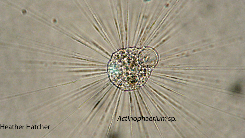
Only observed on gastrotrichia too. He is multicellular and pretty neat looking. He is activly moving but slow enough to get a good look at.
(Smith 2001)
Only observed on gastrotrichia too. He is multicellular and pretty neat looking. He is activly moving but slow enough to get a good look at.
 (Forest 1954)


 (Patterson 2003)
This is the first one of these nemotods that I have seen that didn't squiggle like he was on fire. Most of the time when I would find one he would be moving so fast that the photo was just a blur. But this one seemed to be taking it easy.
(Patterson 2003)
This is the first one of these nemotods that I have seen that didn't squiggle like he was on fire. Most of the time when I would find one he would be moving so fast that the photo was just a blur. But this one seemed to be taking it easy.
 (Smith 2001)
Also known as a Seed Shrimp, he is relitivly large and runs all over the place. He is multicellular and also has a hard shell.
(Smith 2001)
Also known as a Seed Shrimp, he is relitivly large and runs all over the place. He is multicellular and also has a hard shell.
 (Presscott 1964)
This thing barely moved. He is kinda boring.
(Presscott 1964)
This thing barely moved. He is kinda boring.
 (Patterson 2003)
Now the rotifers were plentious and on the move! Found some throughout the glass.
(Patterson 2003)
Now the rotifers were plentious and on the move! Found some throughout the glass.
 (Patterson 2003)
 (Patterson 2003)
On Friday October 26, 2012 ONE Beta Food Pellet was inserted into the microaquarium.
FOOD PELLET INFORMATION:"Atison's Betta Food" made by Ocean Nutrition, Aqua Pet Americas, 3528 West 500 South, Salt Lake City, UT 84104. Ingredients: Fish meal, wheat flower, soy meal, krill meal, minerals, vitamins and preservatives. Analysis: Crude Protein 36%; Crude fat 4.5%; Crude Fiber 3.5%; Moisture 8% and Ash 15%.(McFarland)
Last thursday ( Oct. 25) after I observed my microaquarium, I was instructed to add more water to it from a wash bottle in the lab. I misinterpreted which washbottle to use and used the wrong one. I possibly refilled my microaquarium with 70% ethenal.Eventual, everything in my micro aquarium died, including my plants.


 (Presscott 1964)
So obviously, a new microaquarium had to be made. It has the same type of plants as previously, and the water is from University of Tennessee Hesler Biology Building's greenhouses. After the new microaquarium was made, it was observed. Here are the observations.
 (Patterson 2003)
This little guy above was wiggley and tiny, making it hard to get a good look at him.I saw a few like him and he appears single-celled.
 (Smith 2001)
 (Smith 2001)
 ( Patterson 2003)
 (Smith 2001)
I have only seen one of these. He moves slowly and like a ribbon in the wind. He is multicellular.
 (Smith 2001)
 (Forest 1954)
I didn't zoom in enough to get a good look at them.


I think this mite was dead.
 (Patterson 2003)
 (Smith 2001)
 (Presscott 1964)
 (Patterson 2003)
 (Patterson 2003)
 (Patterson 2003)
(Patterson 2003)
So this microaquarium had alot more life to it. There was more variety as well as quantity .
There were plenty of little tiny unidentifiable organisms as well as a couple of rodifers, about a half dozen epistlelis,a flatworm, a couple of nematodes, about a half dozen colpoda, as well as the organisms pictured above.
Monday, October 29, 2012
Week 1 Observations
Water source :Carter Mill Park at spring source, Carter Mill Road, Knox Co. Tennessee Partial
shade exposure N36 01.168 W83 42.832 940 ft 10/9/2011 ( McFarland)
My Microaquarium !

Observed Thursday, October, 25, 2012
So this week there were a lot more organisms than last week, but still not alot. Things were looking more lively, but some of the water had been depleted.
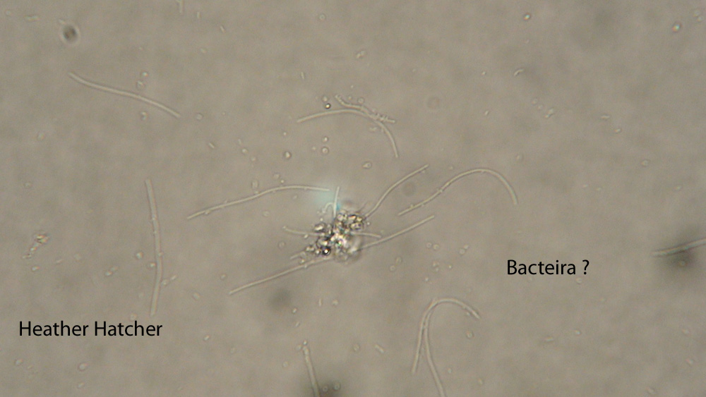
First I observed quite a few organisms that were too small to identify. There were two kinds i kept spotting: long skinning worm like ones, and small black dots running around. The long skinny ones might possibly be bactiera.
Heres is the organisms observed that were large enough to identify and were photogenic, except the Cyclidium sp. , it didnt want its picture taken.
Here's the booger that wouldn't stay still for a picture lol. Why? as his name suggests, he spins around and around in a circluar motion. I saw a couple of organisms like him.
 (Patterson)
 (Patterson)
 (Patterson)
 (Patterson)
 (Patterson)
 (Patterson)

My Microaquarium !

Observed Thursday, October, 25, 2012
So this week there were a lot more organisms than last week, but still not alot. Things were looking more lively, but some of the water had been depleted.
First I observed quite a few organisms that were too small to identify. There were two kinds i kept spotting: long skinning worm like ones, and small black dots running around. The long skinny ones might possibly be bactiera.
Heres is the organisms observed that were large enough to identify and were photogenic, except the Cyclidium sp. , it didnt want its picture taken.
Here's the booger that wouldn't stay still for a picture lol. Why? as his name suggests, he spins around and around in a circluar motion. I saw a couple of organisms like him.
 (Patterson)
I think this next organism is the coolest one I observed.
 (Patterson)
This organism is single celled. He sucks water and other objects into that wide top using two perpellers. Large items hit the perpellers and bounce off in peices. I saw about a dozen of these in my microaquarium. I will have a video up soon.
The next organism is very interesting. I only observed two of them, both pictured below, but one has just eaten something. It's pretty fascinating.
 (Patterson)
The one below has eaten something
 (Patterson)
 (Patterson)
This next organism is pretty cool. It's flagella whips in a circular motion that propels the organism.I only found two of these
 (Patterson)
Here is some of the stuff that was too small to identify. I think it's bactiera. I saw alot of it.
Excited to see next weeks :)
Tuesday, October 23, 2012
Bibliography
McFarland, Kenneth.Botany 111 Fall 2012 Blog
.http://botany1112012.blogspot.com/ .October 21, 2012
Cook R. and McFarland K. General Botany 111 Laboratory
Manual.2012.4th ed. 155-157 p.
Patterson D.J.2003. Free-Living Freshwater Protozoa. London: Manson Publishing Ltd.
Patterson D.J.2003. Free-Living Freshwater Protozoa. London: Manson Publishing Ltd.
Forest, Herman Silva.1954. Handbook of Algae with Special
Reference to Tennessee and the Southeastern United States. Knoxville (TN): The
University of Tennessee Press.
Presscott G.W.1964 The Freshwater Algea.2nd ed.
Dubuque(IA):WM.C Brown Company Publishers.
Smith, Douglas Grant.2001.Pennak’s Freshwater Invertebrates
of the United States: Porifera to Crustcea.4th ed. New York(NY):
John Wiley&Sons,Inc.
Pennak, Robert W.1989.Pennak’s Freshwater
Invertebrates of the United States:Protozoa to Mollusca.3rd ed. New York(NY):
John Wiley&Sons,Inc.
My CORRECTED Post On How I Set-up my Microaquarium.... ( The original post did not contain internal citations)
How did I set up my microaquarium? With help lol.
First I added my water sample to the glass microaquarium
provided. The glass microaquarium has a glass tank, a stand holder, and a lid(Cook
and McFarland). The water sample was placed in the glass tank part. My water
sample came from site #3. Site #3 is Carter Mill Park at spring source, Carter
Mill Road, Knox Co. Tennessee. It is in partial shade exposure. The coordinates
are N36 01.168 W83 42.832 940 ft. It was collected October 10, 2011(McFarland).
Using a pipet, I extracted sediment from the bottom of the container containing
the water sample, and put it inside my glass container first, filling it one
third ways full. Second I extracted water from the sample from about mid-depth,
and placed it in my microaquarium, filling it two third ways full. After that I
extracted surface water from the sample and added it to the microaquarium,
filling the microaquarium almost full. After that I added pieces of two
different aquatic plants to my micro aquarium (Cook and McFarland). The first
plant was Amblestegium sp. Moss. It was collected at the same site as the water
sample(McFarland). The second plant was Utricularia gibba L. It is a flowering,
carnivous plant. The Original material is from south shore of Spain Lake (N
35o55 12.35" W088o20' 47.00), Camp Bella Air Rd. East of Sparta Tn. in
White Co. and grown in water tanks outside of greenhouse at Hesler Biology
Building. The University of Tennessee. Knox Co. Knoxville TN(McFarland). Now my
micro aquarium is set-up!
I placed the lid on the glass microaquarium tank, and
observed its contents under the microscope. I was looking for moving organisms.
After about thirty minutes of observation, with help, I finally found a strand
of pond scum. A little while later a Paramecium was found.
Sunday, October 21, 2012
Citing my entry
Botany 111 Fall 2012 Blog .http://botany1112012.blogspot.com/ .October 21, 2012
Cook R. and McFarland K. General Botany
111 Laboratory Manual.14th ed. 155-157 p.
Setting up my microaquarium !
How did I set up my microaquarium? With help lol.
First I added my water sample to the glass microaquarium
provided. The glass microaquarium has a glass tank, a stand holder, and a lid.
The water sample was placed in the glass tank part. My water sample came from
site #3. Site #3 is Carter Mill Park at spring source, Carter Mill Road, Knox
Co. Tennessee. It is in partial shade exposure. The coordinates are N36 01.168
W83 42.832 940 ft. It was collected October 10, 2011. Using a pipet, I
extracted sediment from the bottom of the container containing the water sample,
and put it inside my glass container first, filling it one third ways full.
Second I extracted water from the sample from about mid-depth, and placed it in
my microaquarium, filling it two third ways full. After that I extracted
surface water from the sample and added it to the microaquarium, filling the
microaquarium almost full. After that I added pieces of two different aquatic
plants to my micro aquarium. The first plant was Amblestegium sp. Moss. It was
collected at the same site as the water sample. . The second plant was Utricularia gibba L. It
is a flowering, carnivous plant. The Original material is from south shore of
Spain Lake (N 35o55 12.35" W088o20' 47.00), Camp Bella Air Rd. East of
Sparta Tn. in White Co. and grown in water tanks outside of greenhouse at
Hesler Biology Building. The University of Tennessee. Knox Co. Knoxville TN.
Now my micro aquarium is set-up!
I placed the lid on the glass microaquarium tank, and
observed its contents under the microscope. I was looking for moving organisms.
After about thirty minutes of observation, with help, I finally found a strand
of pond scum. A little while later a Paramecium was found.
Subscribe to:
Comments (Atom)